
Parking at Southlake
Find Southlake
Donate
Preparing for Care
Frequently Asked Questions

News
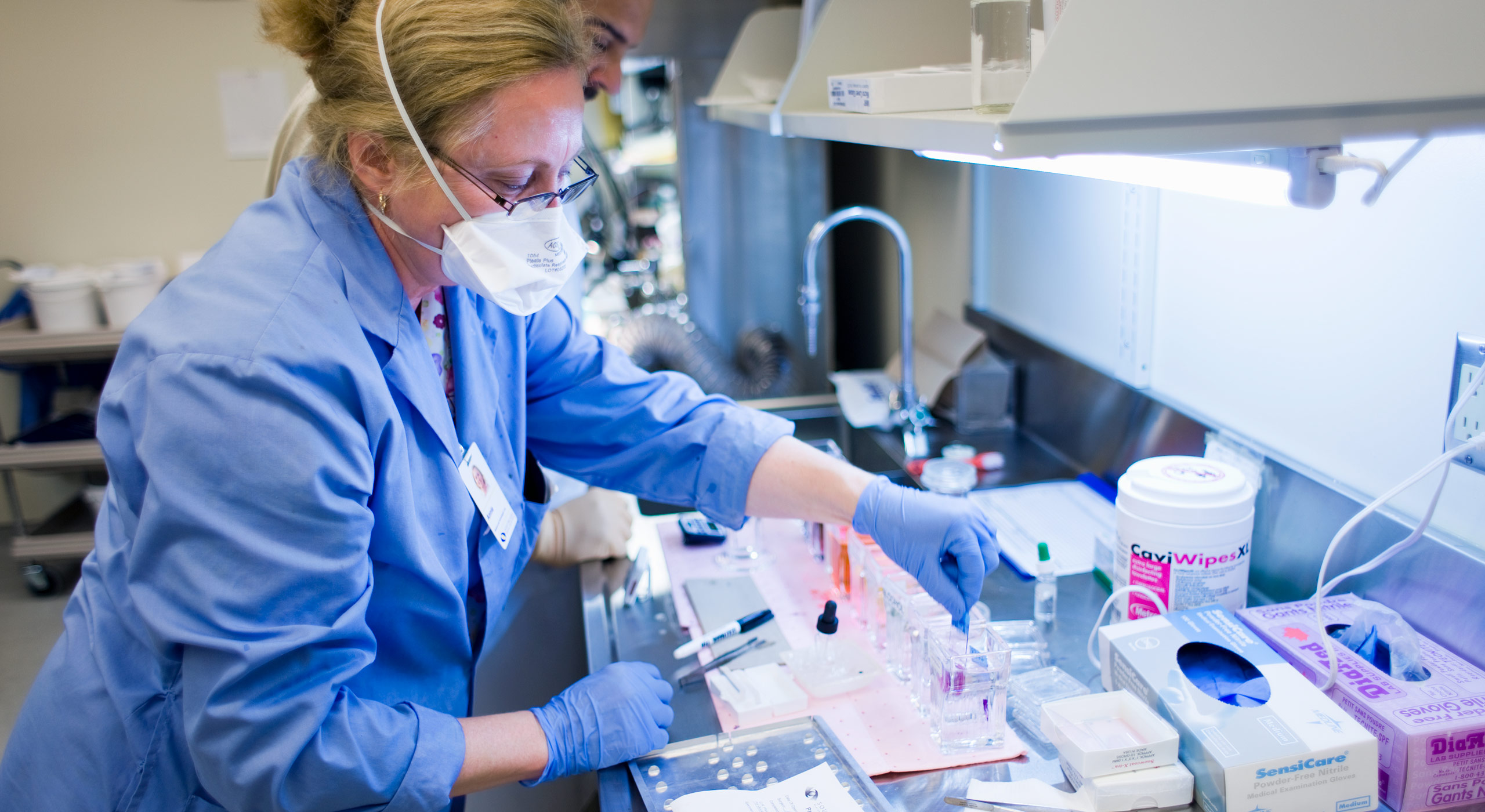
Celebrating National Lab Week
Monday April 12, 2021
Frequently Asked Questions
Bone Marrow Density: How is BMD testing affected by age?
Bone Marrow Density: How is BMD testing affected by age?
If you are younger than 60 years of age and have one or more of the risk factors for osteoporosis, you may want to consider whether BMD testing is right for you. It is possible to reduce your risk for broken bones and other effects of osteoporosis with treatment and/or lifestyle changes. Talk to your physician.
Bone Marrow Density: How much radiation will I be exposed to?
Bone Marrow Density: How much radiation will I be exposed to?
Typically, the exposure for a BMD exam is less than for a standard chest x-ray. As with any x-ray procedure, inform the technologist prior to the exam if there is a chance you may be pregnant.
Bone Marrow Density: What is a Bone Mineral Density (BMD) Exam?
Bone Marrow Density: What is a Bone Mineral Density (BMD) Exam?
Bone Mineral Density (BMD) is a measurement of the concentration of minerals in the bones which are vital for strong bones. A high BMD indicates above average amounts of calcium and phosphates in the bones, while a below-normal bone mineral density can indicate a loss of bone mass, possibly from osteoporosis.
Bone Marrow Density: Who should have a BMD exam?
Bone Marrow Density: Who should have a BMD exam?
The Osteoporosis Society of Canada recommends that people aged 65 or older should have a BMD to screen for osteoporosis. However, if you are at increased risk for osteoporotic fractures, screening should begin earlier.
CT Scan: What are some examples of CT scans when used by physicians in a diagnosis?
CT Scan: What are some examples of CT scans when used by physicians in a diagnosis?
Physicians often use CT scans to:
- Promptly identify injuries to the lungs, heart and blood vessels, liver, spleen, kidneys, bowel, or other internal organs suffering trauma
- Guide biopsies and other procedures (i.e. abscess drainages and/or minimally invasive tumours)
- Determine the results of surgery (i.e. organ transplants and or gastric bypass)
- Set-up and allow for the proper administration of radiation treatments for tumours and/or gauge response from chemotherapy treatment
CT Scan: What is a CT scan?
CT Scan: What is a CT scan?
Computed Tomography or CT scans, combine the use of a computer with a rotating x-ray device to create detailed cross-sectional images or “slices” of the different organs and body sections. These slices are then assembled in two-dimensional, high-resolution images by a computer.
CT Scan: Why is a CT scan unique when imaging for illnesses?
CT Scan: Why is a CT scan unique when imaging for illnesses?
A CT image of the head, for example, allows physicians to also see soft tissue structures, such as the brain and blood vessels. As a result, CT scans provide better precision and reveal more information than a regular x-ray exam.
Interventional Radiology: Is interventional radiology less risky than other tests?
Interventional Radiology: Is interventional radiology less risky than other tests?
While no treatment is risk-free, the risks of interventional radiology procedures are far lower than the risks of open surgery. Most procedures can be performed on an out-patient basis or require only a short stay in hospital.
Interventional Radiology: What is interventional radiology?
Interventional Radiology: What is interventional radiology?
Interventional Radiology is a radiology specialty devoted to the least amount of surgical interruption during your diagnosis and resulting treatment. Our physician radiologists specialize in targeted treatments using image guidance (ultrasound, CT, or x-rays). This helps them to guide small instruments, such as catheters, through blood vessels or through the skin to treat diseases without surgery.
Interventional Radiology: Will I have to go under anesthesia?
Interventional Radiology: Will I have to go under anesthesia?
A general anesthetic is usually not required, but sedation may be offered to keep the patient comfortable during the procedure. Risk, pain, and recovery time are often significantly reduced.
Mammogram: How long does a mammogram take?
Mammogram: How long does a mammogram take?
Your appointment will take approximately 30 minutes to complete.
Mammogram: I’ve heard that getting a mammogram is painful. Is this true?
Mammogram: I’ve heard that getting a mammogram is painful. Is this true?
During the mammogram, each breast will be compressed for only a few seconds. Compression is extremely important as it provides a clearer image of the breast by separating the tissue and also reduces radiation exposure. This should not be painful, although it can be uncomfortable.
Mammogram: Is my mammogram part of the Ontario Breast Screening Program? I’ve heard they are very good at keeping me in touch.
Mammogram: Is my mammogram part of the Ontario Breast Screening Program? I’ve heard they are very good at keeping me in touch.
Yes. Women automatically become part of the Ontario Breast Screen Program (OBSP) when they are referred for a mammogram provided they:
- Are 50 years of age and older
- Have not had previous breast cancer
- Do not have breast implants
- Are not experiencing acute breast symptoms
- Have not had a mammogram within the last year
- Are a current resident of Ontario
The OBSP offers important advantages for women and their primary care practitioners by sending recall and result-letters to women. They also will arrange follow-up services for women if further tests are required. Women who are screened for breast cancer within an organized screening program like the OBSP further benefit by participating in a program that undergoes ongoing quality assurance, program monitoring, and evaluation to ensure that its clients receive high-quality screening.
MRI: Are there risks with getting an MRI?
MRI: Are there risks with getting an MRI?
MRI imaging has no known side effects or risks.
MRI: What are some examples of conditions needing an MRI?
MRI: What are some examples of conditions needing an MRI?
Physicians use MRIs to assist in diagnosing and or monitoring treatment for conditions such as:
- Tumours of the chest, abdomen, or pelvis
- Blockages or enlargements of blood vessels (i.e. aorta, renal arteries and/or arteries in the legs)
- Liver and abdominal disease
- The small intestine, colon and rectum disease
- Cysts and kidney tumours (including other parts of the urinary tract)
MRI: What is an MRI?
MRI: What is an MRI?
Magnetic Resonance Imaging (MRI) is a safe and painless way to take pictures of soft tissues of the body and is especially valuable in diagnosing brain and nervous system disorders, cardiovascular disease, and cancer. An MRI provides greater accuracy in detecting certain diseases and does not require radiation or surgery.
Nuclear Medicine: My doctor has referred me to undergo nuclear medicine testing. Is this safe?
Nuclear Medicine: My doctor has referred me to undergo nuclear medicine testing. Is this safe?
Absolutely. In nuclear medicine, a small amount of radioactive tracer is introduced into the patient to obtain images that show the performance of your internal organs. This radioactive dosage is carefully prescribed to allow for minimal radiation exposure while the patient is positioned under a gamma camera. This camera detects and scans the distribution of the tracer within the patient’s body.
Nuclear Medicine: Will I feel anything when under the camera?
Nuclear Medicine: Will I feel anything when under the camera?
No. You will not feel anything with these drugs and by the following day, most of the radioactive tracer is eliminated from the body by the normal passing of your waste.
Ultrasound: Can I get my results during my ultrasound exam?
Ultrasound: Can I get my results during my ultrasound exam?
No. Your technologist performing the ultrasound cannot provide any results. This includes information regarding the gender of the fetus in pregnant women. Ultrasound information can only be provided by your referring physician.
Ultrasound: Why do I need an ultrasound test?
Ultrasound: Why do I need an ultrasound test?
An ultrasound test can indicate if and when surgery is needed. It can identify and locate aneurysms, blood clots, damaged tissues, heart problems, abnormal growths, and other diseases. It also offers an accurate way to diagnose any fetal abnormalities, multiple pregnancies, tubal pregnancy, cysts, and tumours in the pelvic organs.
What is the benefit of medicine scans?
What is the benefit of medicine scans?
Medicine scans will:
- Analyze how kidneys are functioning and help visualize your blood flow
- Scan patient lungs for respiratory and blood flow problems
- Identify inflammation of the gallbladder and locate an infection
- Evaluate bones for fractures, infection, tumours or arthritis
- Determine cancer presence and to track it from spreading
- Evaluate bleeding of the bowel
- Gauge thyroid function to detect over or underactive thyroids
- Explore abnormalities in the brain and blood flow
- Localize lymph nodes before surgery for patients with melanoma or breast cancer
X-ray: I have an X-ray test scheduled but I feel I may be pregnant. Is this a risk?
X-ray: I have an X-ray test scheduled but I feel I may be pregnant. Is this a risk?
Yes. Radiation has been known to affect the fetus in pregnant women or women who think they may be pregnant. If this is your case, it is advised you should avoid having an x-ray examination. Talk to your physician or technologists if you have any questions about not just an X-ray but any other tests you have been referred for.
X-ray: I’m a little afraid of having an X-ray. Are there any risks?
X-ray: I’m a little afraid of having an X-ray. Are there any risks?
Southlake’s Imaging program uses modern and advanced techniques and equipment which leaves slight risk to radiation exposure. Lead “aprons” and lead shields are used to protect the reproductive organs during most x-ray procedures.